▶지난호에 이어
치근단 질환은 근관을 통해 침투하는 세균에 의해 유발되어 지속되는 것으로써 원인을 제거하기 위해 적절한 protocol에 따라 근관치료를 시행하면 90% 이상의 경우 병소가 치유되고 증상이 소실된다. 그러나 근관치료술식의 진보에도 불구하고 실제 임상에서 근관치료의 성공률은 1950~60년대와 비교하여 그다지 높아지지 않고 있다. 동일한 protocol에 따라 근관치료를 시행한 다음 4~6년 후 환자를 재내원하게 하여 성공과 실패에 영향을 미치는 여러가지 요인들을 관찰하여 보고한 Toronto study에 의하면 통상적인 근관치료를 시행한 경우 86%의 성공률을 보였고4-7), Strindberg가 1956년에 보고한 성공률과 비교해 볼 때 그다지 차이가 없다.8)
근관형성, 근관세척 후에도 근관내 세균은 어느 수준까지는 감소하지만 치근단에서는 거의 세균이 존재하지 않는다. 이후 근관을 3차원적으로 충전하면 치근단의 면역기능에 의해 치근단 질환은 치유된다. 그럼에도 근관치료가 실패하게 되는 첫째 원인으로 근관계의 치아별 복잡한 형태와 해부학적 변이, missing canal 등에서 찾아볼 수 있다(그림 1, 2, 3, 4, 5). 둘째는 Apical delta, 측방관(lateral canal) 등 근관치료 술식으로 도달되지 않는 곳에서 존재하던 세균이 지속적인 감염의 원인이 되고, 시간이 지나 근관충전물의 미세누출이 생기면 치근단염을 유발할 수 있다12)(그림 6, 7, 8, 표 8, 9). 셋째, 근관치료 후 최종 보철로 수복이 되지 않을 경우 coronal leakage에 의하여 2차 감염이 발생할 수 있다. 넷째, 만성 치주염, 확인되지 않은 Crack도 근관치료 실패의 원인으로 작용한다.

그림 1. Micro-.computed tomographic scans of dental anatomy (36μm resolution)
A, Clinical view of tooth #21 shows two accessory canals and an apical bifurcation.
B, Mesiodistal view of the tooth shown in A.
C, Working length radiograph, with files placed in both apical canal aspects(Pathway of the pulp, 10th, 293)
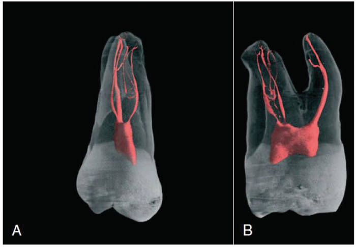 그림 2. Micro.computed tomographic scans of more complicated dental anatomy (36 μm resolution).
A, Clinical view of tooth #16 shows a fine mesiobuccal and distobuccal canal system with additional anatomy in all three roots.
B, Mesiodistal view of the tooth shown in A.(Pathway of the pulp, 10th, 293)
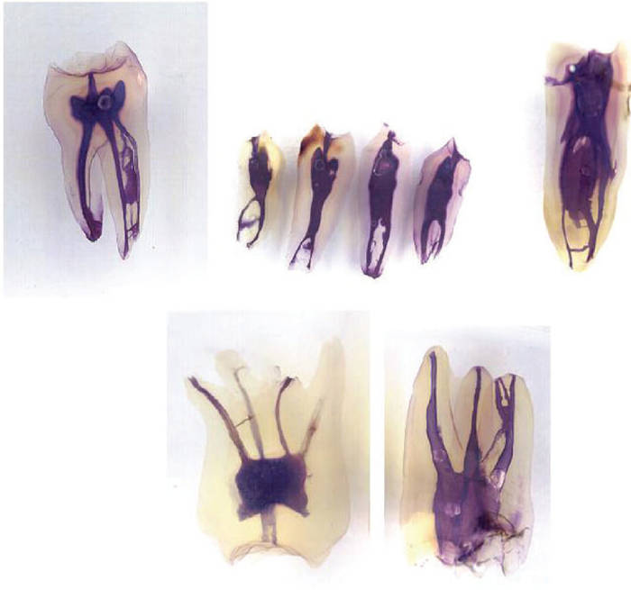 그림 3. Cleared teeth demonstrating root canal variation (FRANK J. VERTUCCI, Root canal morphology and its relationship to endodontic procedures. Endodontic Topics 2005, 10, 3-9)
(A) Mandibular second molar with three mesial canals.
(B) Mandibular premolars with Vertucci type V canal configuration.
(C) Mandibular premolars with three canals and intercanal connections.
(D) Maxillary second molar with two palatal canals.
(E) Maxillary first molar with two canals separating into three in mesiobuccal root. MB-2 orifice close to palatal orifice.
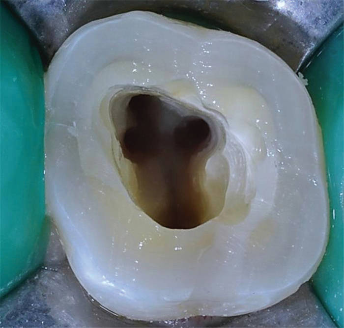
그림 4. 상악 제1대구치의 MB2가 관찰되는 4근관, 박용훈 원장(메디힐치과) 제공
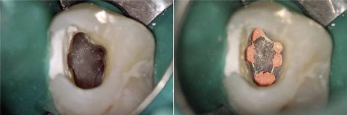
그림 5. 5개의 근관을 가진 상악 제1대구치
(Martins et al, Endodontic treatment of the maxillary first molar with five root canals-Three case reports, Rev Port Estomatol Med Dent Cir Maxilofac. 2013;54:37-42)
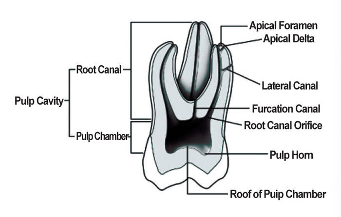
그림 6. Major anatomic components of the root canal system
(FRANK J. VERTUCCI, Root canal morphology and its relationship to endodontic procedures. Endodontic Topics 2005, 10, 3-9)
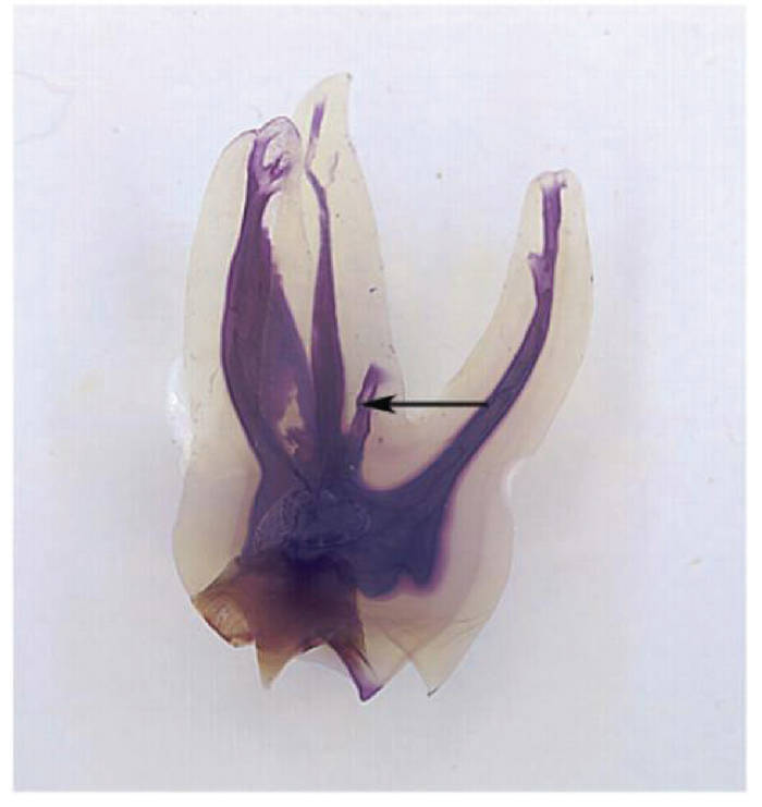
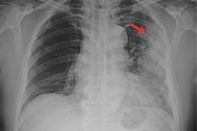
그림 7. Maxillary first molar illustrating a furcation canal(arrow)
(FRANK J. VERTUCCI, Root canal morphology and its relationship to endodontic procedures. Endodontic Topics 2005, 10, 3-9)
4) Friedman S, Abitbol S, Lawrence HP. Treatment outcome in endodontics : The Toronto study. Phase 1: initial treatment. J Endod 2003. 29:787-793,
5) Farzaneh M, Abitbol S, Lawrence HP, Friedman S. Treatment outcome in endodontics : The Toronto study. Phase II: initial treatment. J Endod 2004. 30:302-309,
6) Marquis VL, Dao T, Farzaneh M, Abitbol S, Friedman S. Treatment outcome in endodontics : The Toronto study. Phase III: initial treatment. J Endod 2006. 32:299-306,
7) de Chevigny C, Dao TT, Basrani BR, Marquis VL,Farzaneh M, Abitbol S, Friedman S. Treatment outcome in endodontics : The Toronto study. Phase 4: initial treatment. J Endod 2008.34:258-263,
8) Strindberg LZ. The dependence of the results of pulp therapy on certain factors. An analytic study based on radiographic and clinical follow-up examinations. ActaOdontol Scand 1956 14:1-175,
9) Cohen, Pathway of the pulp, 10th
10) FRANK J. VERTUCCI, Root canal morphology and its relationship to endodonticprocedures. Endodontic Topics 2005, 10, 3?9)
11) Martins et al, Endodontic treatment of the maxillary first molar with five root canals ? Three case reports, Rev Port Estomatol Med Dent Cir Maxilofac. 2013;54:37-42)
12) Walton & Torabinejad, Principles and practice of endodontics 1989